Clinical Topic: Ethanol intoxication, Brain injury and outcome

All CRNAs who take call in a medical center have had the challenge of caring for the intoxicated driver who arrives at the Hospital with a head injury.  Imagine that both drivers sustained a traumatic head injury.  One driver was intoxicated and the other was completely sober.  Which driver is more likely to have a better outcome?

A study by Lustenberger, T, et al published in the Journal of Neurotrauma looked at outcomes of patients with traumatic brain injury. The aim of this study was to determine the impact of ethanol (ETOH) on the incidence of severe traumatic brain injury associated coagulopathy and to examine the effect of ETOH on in-hospital outcomes in patients sustaining sTBI.  The incidence of admission coagulopathy, in-hospital complications, and mortality were compared between patients who were ETOH positive and ETOH negative.

The authors found that coagulopathy was significantly less frequent in the ETOH (+) patients compared to their ETOH (-) counterparts

For brain-injured patients arriving alive to the hospital, ETOH intoxication is associated with a significantly lower incidence of early coagulopathy and in-hospital mortality.

Click here to read an abstract and link to the original article by Thomas Lustenberger.

CRNA Fitness: Nutrition

The Gas Range
By Liz Sanner Davis

Think of the food on your plate as Fuel.  Think of the container it comes in as the Gas-can.  And label the inside of your body as The Tank.  At every meal you must carry the fuel in a gas can and deliver it unpolluted to the tank if you want to have enough gas to live life and to give life in the OR.  When the patient is short on gas, he crumps.  When you are short on gas, you crump.  And when low-grade gas is delivered to the tank ( think  paper bags with famous logos, cardboard boxes with grease-covered bottoms, and paper cups with sippy devices), you and your patient may both wind up driving on empty.  During the month of January, try making this change in your nutrition habits at work.  Bring fresh lunch foods to work prepared in your own kitchen – last night’s leftover salmon is a worthy protein, light yogurt and low-sugar granola, a crisp apple, a ripe avocado big enough to share are all perfect pick-me-ups. Or stack your own turkey sandwich made with whole grain bread, lettuce and tomato.  Avoid saltyfoods from the cafeteria and resist the urge to send out Smiling Samantha for pepperoni pizza with stuffed cheese crust, “double-the-olives.”  Fill your tank with superior grade fuel delivered in a pollutant-free gas-can.  You’ll sustain two lives – yours and the patient’s.

Liz Sanner Davis is owner and trainer at Body Firm Integrated Fitness Solutions, Temple, TX.  Visit Liz online at www.bdyfrm.com and read her humorous and motivational Lizlines or Lizlimericks published daily.

Radiation Exposure to Anesthetists

Radiation exposure has long been a concern to Radiologists and their assistants.  Lead aprons, thyroid shields and more recently leaded eyeware have reduced their exposure to radiation.  Anesthetists are frequently assigned to sedate / anesthetize patients in the interventional radiology suite.  Like the Radiologist, the anesthetist is exposed to radiation danger.

In a study by Anastasian, ZH, et al published in Anesthesiology, the facial exposure to radiation was compared between the Radiologist and the Anesthetist.   Both providers wore lead aprons.   The Radiologist wore leaded eyeglasses and stayed behind a leaded acrylic shield to the extent possible.  The Anesthesiologist also was instructed to stay behind a leaded shield to the extent possible and to keep maximum distance from the source of radiation.

The authors of the study demonstrated that the Anesthesiologist had a 3 fold increase in facial exposure to radiation than did the Radiologist, thus increasing the risk of developing cataracts.  Exposure of the Anesthesiologist was correlated to the number of pharmacologic interventions performed during the case.

The best ways to reduce exposure to radiation are distance and shielding.  Lightweight leaded eyeglasses reduce exposure of the cornea by 98% and are recommended for those spending significant time administering anesthesia for interventional radiology

Click here to review the work by Anastasian, ZH et al.

Please return to this site and leave a comment

Opioid-Induced Respiratory Depression

The Anesthesia Patient Safety Foundation is committed to the safe and reliable administration of every anesthetic.  An area of concern has been respiratory depression in the immediate postoperative period secondary to narcotic administration.  The following information comes directly from the foundation;

The APSF believes that clinically significant, drug-induced respiratory depression in the postoperative period remains a serious patient safety risk that continues to be associated with significant morbidity and mortality since it was first addressed by the APSF in 2006.1 The APSF envisions that “no patient shall be harmed by opioid-induced respiratory depression in the postoperative period,” and convened the second multidisciplinary conference on this serious patient safety issue in June of this year in Phoenix, AZ, with 136 stakeholders in attendance. The conference addressed “Essential Monitoring Strategies to Detect Clinically Significant Drug-Induced Respiratory Depression in the Postoperative Period.”

Click here to read the full report by the APSF regarding drug-induced depression in the postoperative period.  After reading the report, click the back arrow to return to this site and leave a comment for your colleagues.

Ketamine Supplement for Anesthesia

Ketamine is an old drug which has come in and out of favor many times over the past decades.  Currently, articles are emerging touting the advantages of Ketamine as a supplement for  both general anesthesia and monitored anesthesia care.  Below is a description by Pamela Chambers, CRNA regarding her clinical experience using Ketamine as a supplement.  Read her excellent review, scan the articles and leave a comment to share your experiences with your colleagues.

Submitted by Pamela Chambers, CRNA

Lexterrae legal consulting service

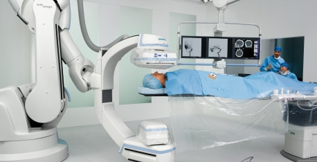
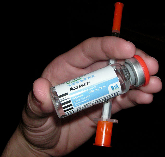
For the EP case that lasted 6+ hours, GETA with .5 mac Desflurane and Propofol infusion at 30 ug/kg/min, the Propofol Ketamine mixture performed very well.  I mixed 50mg Ketamine in each 50ml bottle of propofol.  I used less than 3 bottles for the case and never used more than .5 mac Des.  I used a total of 25 ug Fent, just after intubation, for the case.  The patient was 83 yo male, approx 90 kg, EF 20%.

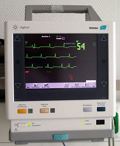
Upon emergence the patient denied any pain or discomfort.  He was appropriately responsive to verbal and did not cough on extubation. VS were stable and he was A&O x3 on arrival to the EP room for phase 2 recovery.

A few days later, I used Ketamine as an analgesic adjunct for another long case (10 hr bilateral mastectomy and tram flap).  I used a total of 195 mg Ketamine.  The patient was an ASA 1.  After 100 ug Fent (and 3mg Versed) prior to induction, I used 30 mg Ketamine approximately 3 mins prior to incision.  Then I administered 30 mg Ketamine at hr number 2, and hr #3.  Around hr #4, I decreased the Ketamine to 15mg for each hrs successive dose with a plan to halt Ketamine admin when I believed the case was 2 hrs from completion.  My last Ketamine dose was at 1430 during the last phases of the case. The patient began spontaneous ventilations after reversal of NDMR at approximately 1640.  The case ended at approximately 1730.  The patient

received a total of 2200 ug Fent dosed approximately 100 ug every 30-45 mins.

Intermittent rescue doses for SNS spike were not required. Pressor agents were not used. Hemodynamics were extremely stable, almost like the proverbial railroad track!

Total fluid administered was 2 liters NS and 1 liter 5% Albumin, UO was 150 ml, EBL 300ml. I also adminstered 2 mg Versed at 1600 to facilitate decreasing the inhaled agent level and to ameliorate any emergence delirium associated with the Ketamine.  The patient was appropriately responsive to verbal prior to extubation, and did not cough during extubation.  She also denied any c/o pain immediately post-operatively. No emergence delirium was appreciated.

Click here to read a review article by Laskowshi et. al regarding Ketamine use

Click here to read an article by McCartney et al regarding the role of the NMDA receptor
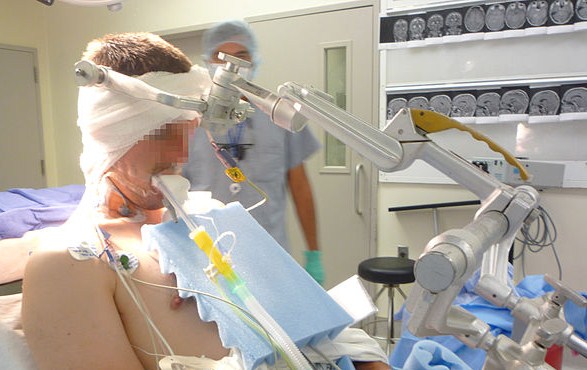
LMA in the Prone Position

The LMA (Laryngeal Mask Airway) has been a common airway management device used by the Anesthesia community for two decades.  Because the device does not “secure” the airway like a cuffed endotracheal tube would, anesthetists are selective about the patient population and type of surgery when deciding whether or not to use the LMA.  Our international colleagues seem to have been bolder with the use of the LMA and report its use in the lateral and sitting positions.   An Article by Ng Published in Anesthesia and Analgesia reported a series of over 200 patients who were safely induced in the prone position with the LMA inserted after induction.  Click here to read the article and then return to procrna.com and leave a comment.  Let us know about your experience using the LMA in the non-supine patient.